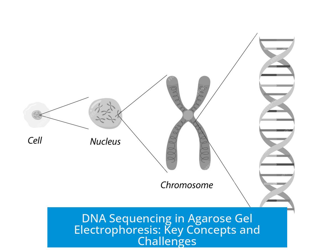
DNA Sequencing in Agarose Gel Electrophoresis
DNA sequencing in agarose gel electrophoresis involves using gel-based separation of DNA fragments to analyze plasmid DNA conformations and assess digestion patterns prior to sequencing. This process helps verify DNA integrity and mutation-induced changes before sequencing steps.
Understanding DNA Bands in Agarose Gel
Agarose gel electrophoresis separates plasmid DNA into distinct band forms:
- Supercoiled DNA: The most compact form runs fastest in the gel, appearing as the lowest band. It binds more ethidium bromide and fluoresces more intensely due to its density.
- Open Circular DNA: The relaxed form moves slower, showing as the upper band. Nicked or ‘nicked lariats’ often migrate slower than expected, sometimes appearing larger on the gel.
- Linear DNA: Resulting from complete or partial digestion, the linear form typically runs between supercoiled and nicked forms. For example, a single cut plasmid of ~7 kb shows a sharp linear band.
DNA Ladder Usage and Interpretation
DNA ladders provide size references for estimating fragment lengths.
- Ladder bands are marked with known sizes, which enable comparison to sample bands.
- It is important to note that ladders cannot accurately size undigested plasmids since different plasmid conformations migrate irregularly.
- High-quality ladder bands help clarify sample interpretation but do not resolve plasmid conformation ambiguity.
Challenges in Band Size Interpretation
Identifying plasmid band types and sizes presents challenges:
- Supercoiled, nicked, and linear DNA differ in gel mobility unpredictably.
- Mutation sites, such as introduced AseI sites, may alter digestion patterns, creating linear bands detectable by gel electrophoresis.
- Distinguishing between bands based solely on migration requires experience, especially when samples include partial digests or mixed conformations.
Role of Gel Electrophoresis in DNA Sequencing
Prior to DNA sequencing, agarose gel electrophoresis confirms plasmid preparation quality:
- Ensures plasmid purity by identifying unwanted forms or degradation.
- Verifies complete digestion at introduced mutation sites to guide sequencing primer design.
- Assesses DNA quantity and fluorescence intensity related to conformation for efficient sequencing.
In some cases, plasmids are cut to linearize DNA for sequencing reactions, improving read accuracy.
Summary
- Supercoiled plasmids run faster and fluoresce brighter on agarose gels.
- Open circular and nicked plasmids exhibit slower migration and appear as higher bands.
- DNA ladders assist in size estimation but cannot size uncut plasmids precisely.
- Gel electrophoresis reveals digestion efficiency and integrity before sequencing.
- Plasmid mutations can alter digestion patterns, detectable as linear bands.
What do different plasmid DNA bands represent in agarose gel electrophoresis?
Supercoiled DNA forms the bottom band and runs faster. Open circular DNA appears as the top band. Nicked lariats look larger than their actual size and migrate slower than supercoiled DNA.
Why does supercoiled DNA appear brighter after ethidium bromide staining?
Supercoiled DNA binds more ethidium dye due to its dense structure. This causes it to fluoresce more brightly than other forms like nicked or linear DNA.
Can you determine plasmid size using an uncut DNA ladder?
No, ladders cannot estimate plasmid size if the plasmid is uncut. Only linearized DNA fragments or digested plasmids correlate properly with ladder markers.
How does partial digestion affect plasmid band patterns?
Partial digestion creates linear DNA bands, often near 7 kb for a single-cutter site. This linearized plasmid runs differently than supercoiled or nicked forms.
Why is it hard to interpret bands when combining ladder and different plasmid forms?
Ladder bands mark known sizes, but plasmid forms (nicked, supercoiled, linear) run differently. This makes direct size comparison with ladders complex and sometimes confusing.
Leave a Comment