Understanding GFP (Green Fluorescent Protein)
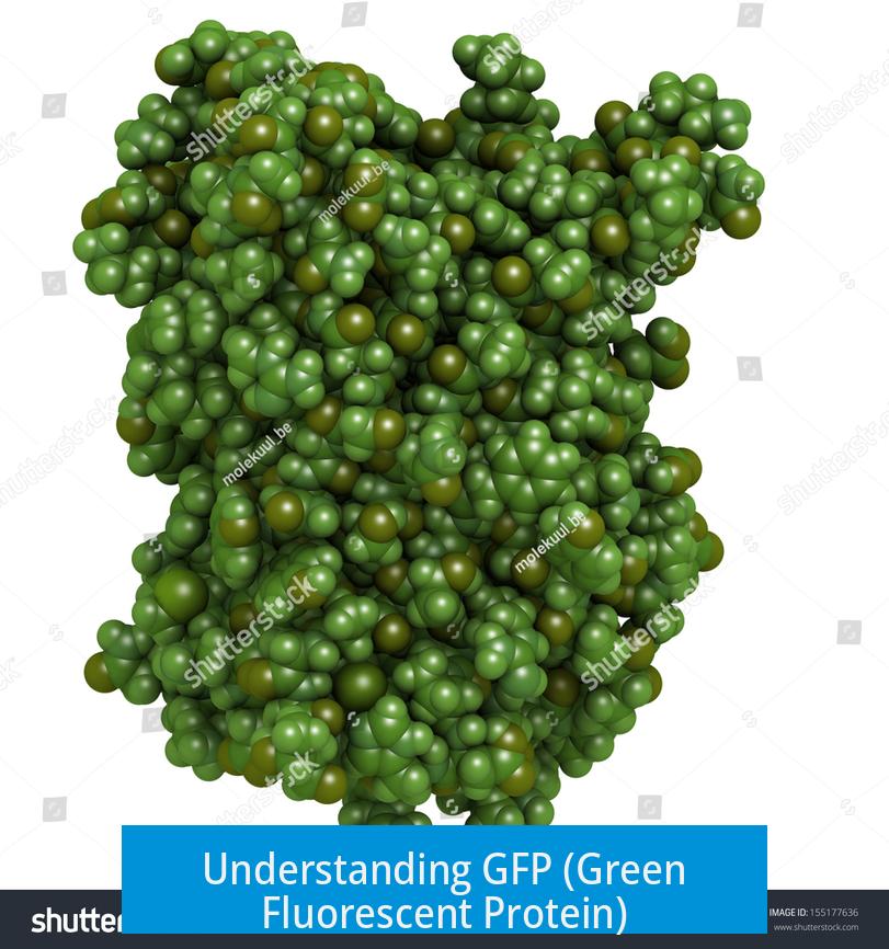
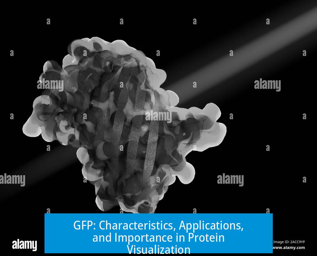
GFP is a protein that glows brightly when exposed to specific light, allowing visualization of proteins inside living cells. This glowing property makes GFP an invaluable tool in molecular biology for tracking and studying proteins in real time.
Basic Characteristics and Function
GFP naturally fluoresces green under blue to ultraviolet light. It is classified as a reporter protein because it signals the presence of specific proteins by emitting light. When linked to other proteins, GFP “reports” their expression by glowing, making these proteins visible under a microscope.
Gene Expression and Fusion with GFP
Researchers fuse GFP to a gene of interest, placing it near a promoter region or coding sequence. This fusion means the gene and GFP are expressed together. If the gene is active, GFP glows, indicating protein production.
- Placing GFP downstream of a promoter shows if and when a gene is activated.
- Fusing GFP at the end of a coding sequence reveals how much protein is produced.
- Applicable to any gene, from those correcting mutations to those producing functional proteins.
Visualization and Tracking of Protein Localization
GFP allows researchers to see exactly where a protein localizes in the cell, aiding in understanding protein function. Under normal light microscopy, cellular structures appear vague. GFP tagging highlights specific proteins, clarifying their location.
- Identifies unknown cellular structures by correlating glowing specks with known proteins.
- Tracks protein migration within the cell over time.
- Reveals the site of protein actions during cellular processes.
Stability and Dynamics of GFP
Cytosolic GFP has a half-life slightly over 24 hours, allowing observation of protein dynamics within a meaningful timeframe. Changes in protein levels or localization can be monitored as cells live and react.
Practical Applications and Impact
GFP helps define protein roles by indicating presence or absence of fluorescence. It also can enhance protein expression levels when fused. The reliability and versatility of GFP earned it a Nobel Prize, recognizing its transformative impact on life sciences.
Key Takeaways
- GFP glows under specific light, enabling protein visualization.
- It functions as a reporter by fusing with genes to trace expression.
- GFP reveals protein location and dynamics inside living cells.
- Its stability allows monitoring over extended periods.
- GFP’s application revolutionized biological research and won a Nobel Prize.
What makes GFP glow under a microscope?
GFP emits bright light when exposed to a specific wavelength. This glowing property allows it to be seen clearly inside cells.
How does fusing GFP to a gene help in research?
By fusing GFP to a gene, researchers track if that gene is expressed. If the fused GFP lights up, it confirms the gene is active and producing protein.
Can GFP show where a protein is located inside a cell?
Yes. GFP tags a protein to reveal its exact location within the cell. This helps identify structures and understand the protein’s function.
What is the significance of GFP’s half-life?
The half-life of GFP in the cytosol is about 24 hours. This stability allows observation of protein dynamics within a meaningful timeframe.
Does attaching GFP affect the protein it marks?
Sometimes GFP tagging can increase protein expression. It may also aid in detecting the protein more easily during experiments.


Leave a Comment