Difference Between Loading Dye and Gel Stain in Electrophoresis
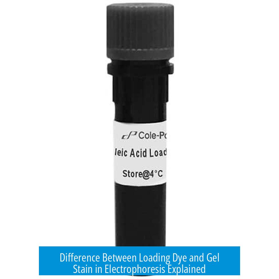
The main difference between loading dye and gel stain lies in their roles during electrophoresis: loading dye is mixed with the DNA samples before loading into wells to weigh down the DNA and provide visual tracking, while gel stain is incorporated into the gel or applied afterward to bind DNA and enable visualization under UV light.
Loading Dye: Function and Use
Loading dye is added directly to the DNA sample before electrophoresis. It has two key functions:
- Weights down DNA: It increases the density of the sample. This prevents the DNA from floating out of the wells when placed in the gel.
- Visual tracking during run: The dye contains colored tracking molecules that migrate through the gel at predictable rates. This helps monitor the progress of electrophoresis in real-time, so the run can be stopped before DNA runs off the gel.
This mixture is simple and quick to apply, such as using 1 μL of an orange-blue loading dye per 25 μL reaction. It does not stain or bind DNA permanently but facilitates loading and monitoring.
Gel Stain: Function and Use
Gel stain is a DNA-binding dye used for post-run visualization. It binds to nucleic acids and fluoresces when exposed to ultraviolet (UV) light.
- Incorporated in molten gel: The stain can be mixed into the molten agarose before casting, enabling uniform staining.
- Used after electrophoresis: Alternatively, the gel can be soaked in a staining solution, such as 10 μL Promega nucleic acid stain in 35 mL of 1x TAE buffer for 30 minutes, to stain DNA bands.
- Facilitates imaging: Fluorescing under UV light, the gel stain allows visualization and documentation of DNA band locations with imaging systems.
Summary Table
| Aspect | Loading Dye | Gel Stain |
|---|---|---|
| Added To | DNA sample before loading | Molten gel or post-run gel |
| Primary Purpose | Weight sample, track progress | Visualize DNA bands post-run |
| Visible During | During electrophoresis | After electrophoresis under UV |
| Effect on DNA | Does not bind DNA permanently | Binds DNA and fluoresces |
Key Takeaways
- Loading dye helps load samples and visually track DNA migration during electrophoresis.
- Gel stain binds to DNA to allow imaging after electrophoresis via fluorescence under UV light.
- Loading dye affects sample density; gel stain affects visualization sensitivity.
- Both are essential but serve distinct purposes in gel electrophoresis workflows.
What is the main function of loading dye in electrophoresis?
Loading dye is mixed with your DNA sample to weigh it down, preventing it from floating out of the wells. It also provides a visible marker to track how far the DNA has moved during electrophoresis.
How does gel stain differ in its application compared to loading dye?
Gel stain is mixed directly into the molten agarose gel before it solidifies. Unlike loading dye, it binds to DNA within the gel, allowing visualization under UV light after the run.
Can loading dye be used to visualize DNA bands after electrophoresis?
No, loading dye only helps track DNA migration during the run. For visualizing DNA bands after electrophoresis, gel stain is used since it fluoresces under UV light.
Why is gel stain important for imaging DNA on the gel?
Gel stain binds to the DNA and fluoresces, enabling imaging machines to detect and record the precise location and intensity of DNA bands on the gel.


Leave a Comment