Why Is LC3 II Upregulated When Autophagy Is Inhibited?
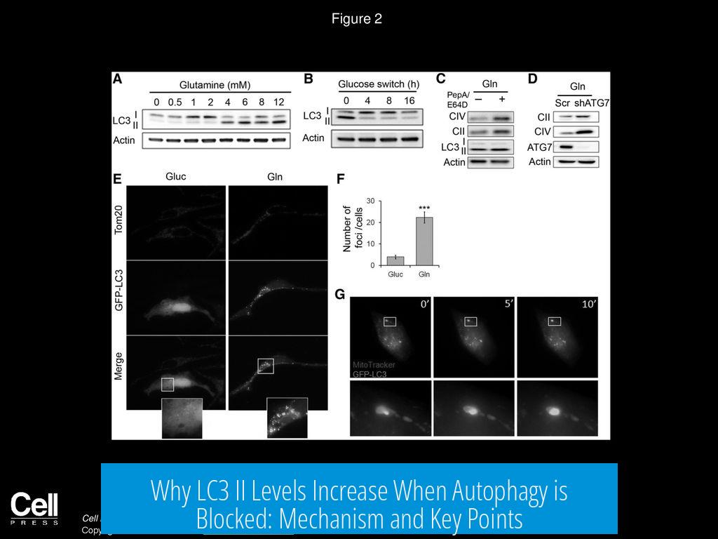
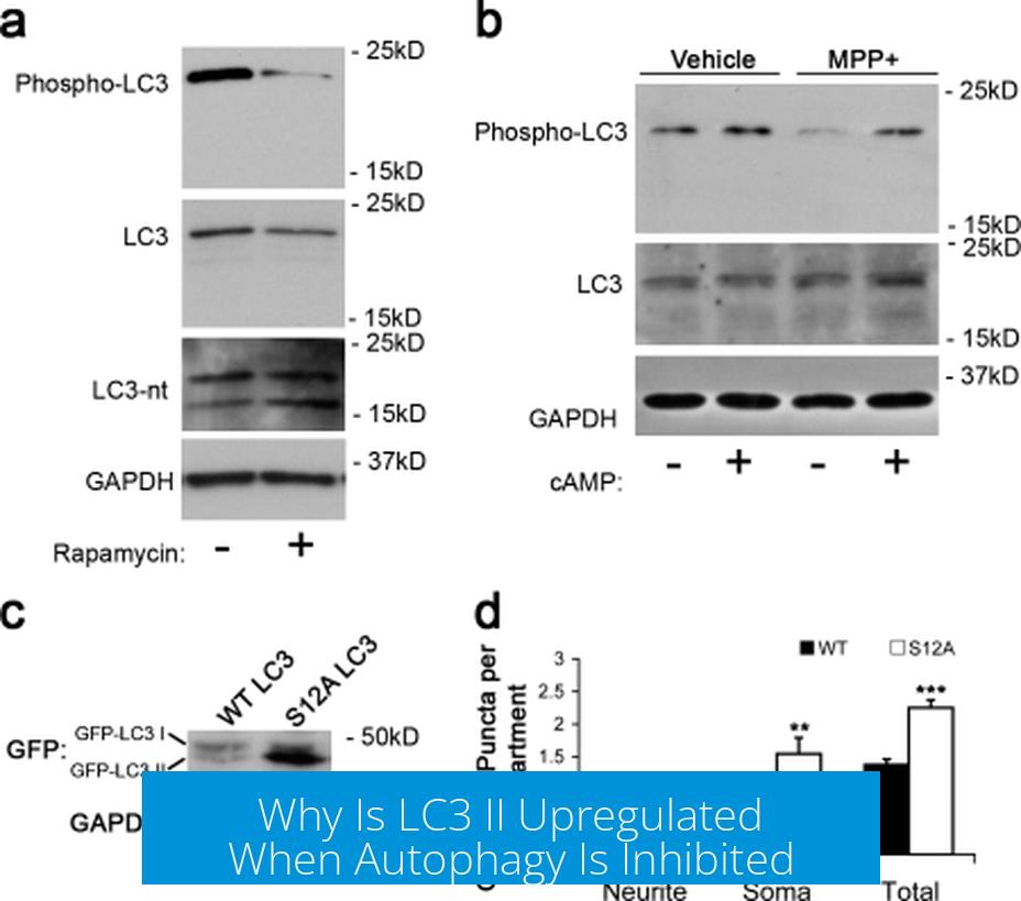 LC3 II is upregulated when autophagy is inhibited because the final degradation step of autophagy is blocked, causing LC3 II-containing autophagosomes to accumulate in the cytoplasm. This occurs due to the failure of autophagosomes to fuse with or be degraded in autolysosomes.
Mechanism of LC3 II Upregulation
LC3 (microtubule-associated protein 1 light chain 3) exists in two forms: LC3 I (cytosolic) and LC3 II (membrane-bound). LC3 II associates with autophagosomal membranes during autophagy. Normally, autophagosomes mature by fusing with lysosomes to form autolysosomes where the contents, including LC3 II, are degraded.
When autophagy inhibitors such as bafilomycin A1 or chloroquine are applied, they block the fusion of autophagosomes with lysosomes or alter lysosomal pH. This inhibits the final degradation step. As a result, LC3 II-labeled autophagosomes cannot be cleared and accumulate in the cytoplasm, causing an observable increase in LC3 II levels.
LC3 II as a Marker of Autophagy Flux
Measuring LC3 II levels after autophagy inhibition reveals the flux, or activity, of the autophagy pathway under normal conditions. Since autophagosomes accumulate only when degradation is blocked, higher LC3 II after treatment reflects more active autophagy prior to inhibition.
- Cells with high autophagic flux produce more autophagosomes, leading to greater LC3 II buildup when degradation stops.
- Comparing LC3 II increase among different cell lines after inhibition can indicate differences in autophagy rates.
For example, wild-type cells may show a larger LC3 II increase upon bafilomycin treatment than knockout cells lacking autophagy genes. This supports that wild-type cells have higher autophagic activity.
Summary of Key Points
- LC3 II upregulation occurs because autophagosomes cannot be degraded when autophagy is inhibited.
- Inhibitors block autophagosome-lysosome fusion or lysosomal function, causing LC3 II accumulation.
- Measuring LC3 II levels after inhibition estimates autophagy flux in cells.
- Greater LC3 II increase indicates higher baseline autophagic activity.
- Comparisons among cell lines after inhibition provide insight into their autophagy efficiency.
Why does LC3 II increase when autophagy is blocked?
LC3 II builds up because drugs like bafilomycin block the final autophagy step. Autophagosomes with LC3 II cannot be degraded and accumulate in the cytoplasm.
How does LC3 II accumulation reflect autophagy flux?
The rise in LC3 II after inhibition shows how active autophagy was. More LC3 II means more autophagosomes formed before the block.
Can LC3 II levels compare autophagy in different cells?
Yes. Cells with higher LC3 II after treatment have greater autophagy flux. Comparing levels reveals differences in autophagosome turnover between cells.
What role do autolysosomes play in LC3 II regulation?
Autolysosomes degrade autophagosomes with LC3 II. When fusion or degradation is blocked, LC3 II cannot be removed and accumulates.
Is LC3 II accumulation a direct measure of autophagy inhibition?
It indicates blockage in autophagosome degradation but not complete autophagy inhibition. Accumulation results from halted terminal steps rather than ceased autophagosome formation.


Leave a Comment